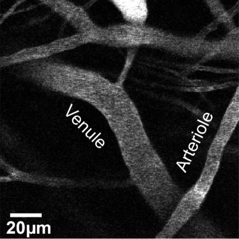
In vivo two photon image of dextran labeled blood vessels in an experimental model
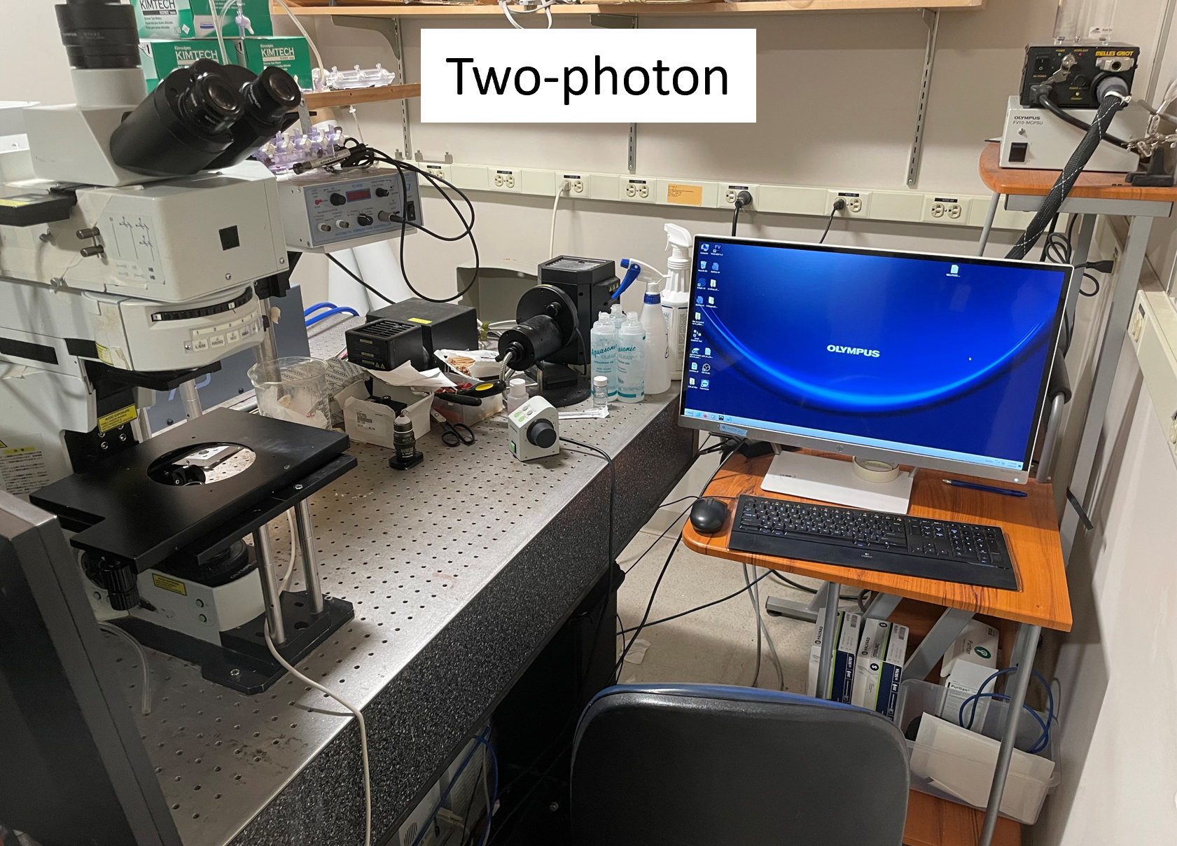
Our favorite microscope
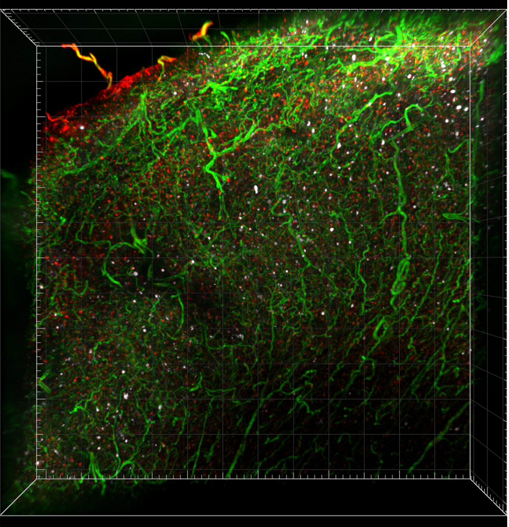
Confocal image of a 1 mm thick piece of cleared cortical tissue with blood vessels (green), tau tangles (white), and neurons (red) labeled.
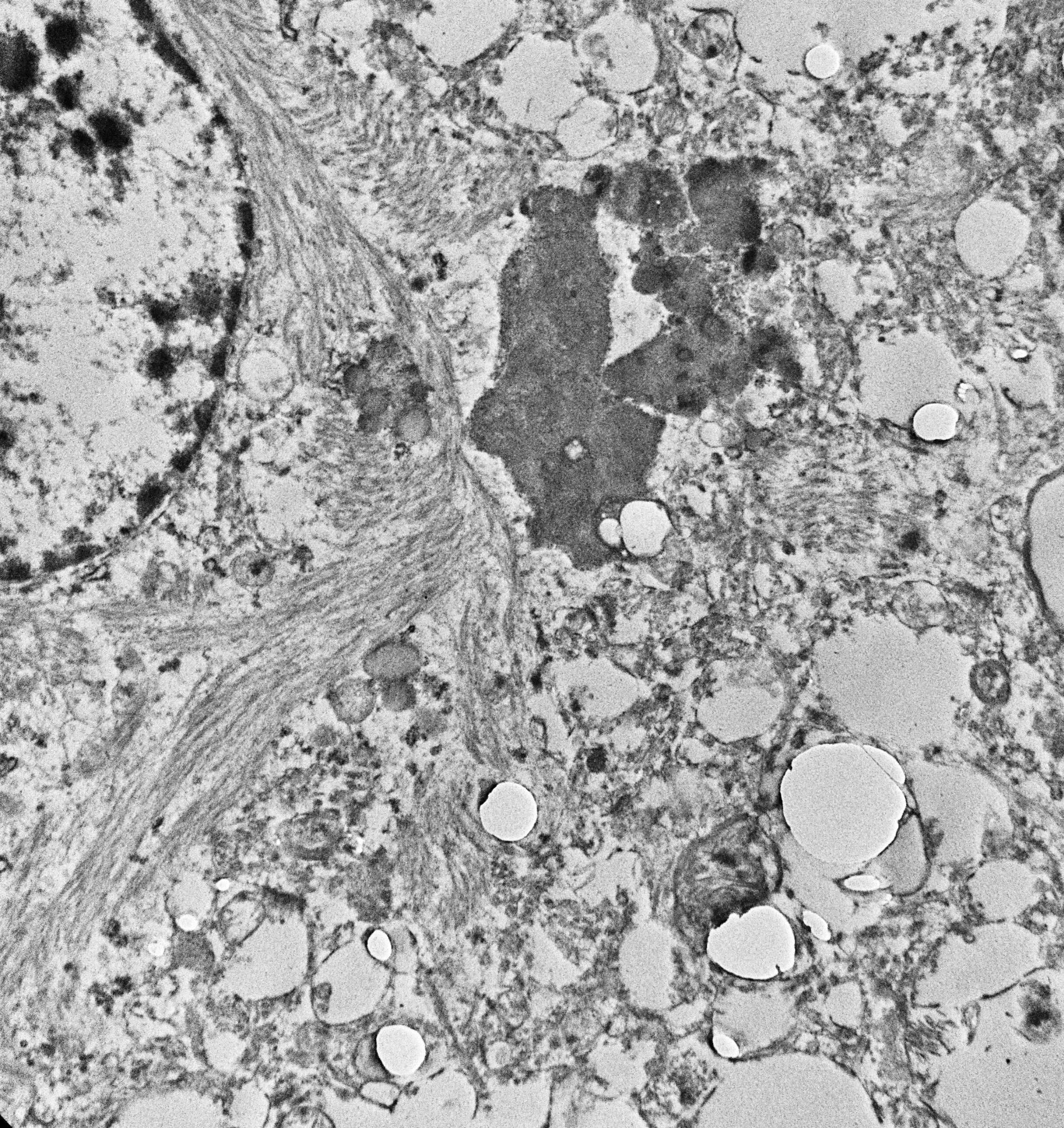
Electron micrograph image of a tangle-bearing neuron in an AD brain
View of the lab
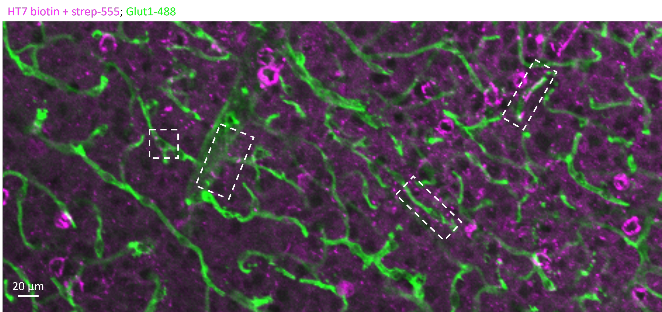
Tau-containing neurites and tangles near blood vessels
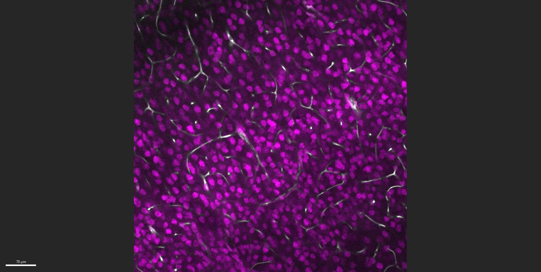
Glut1-labeled blood vessels (white) and NeuN-labeled neurons (magenta)
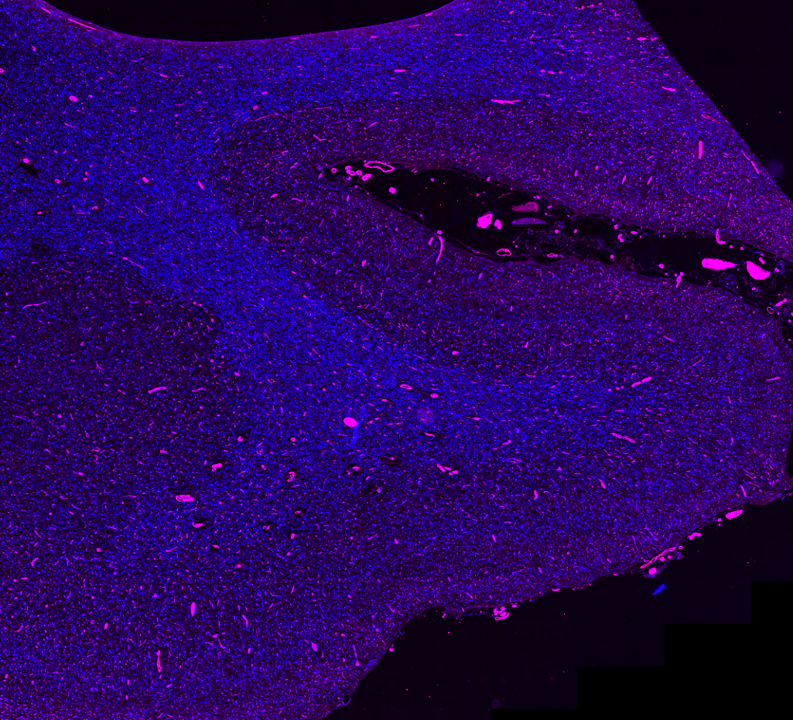
Temporal cortex from AD brain labeled for blood vessels (magenta) and cell nuclei (blue)